ADVERTISEMENTS:
In this article we will discuss about the vegetative and sexual modes of reproduction in Marchantia.
Vegetative Reproduction in Marchantia:
Vegetative propagation of Marchantia takes place by the following methods:
(a) By progressive Death and Decay of the Thallus:
ADVERTISEMENTS:
Like Riccia, the vegetative propagation of Marchantia is quite common which takes place by the unlimited growth of the branches and decay from the base. When the decay of cells reaches dichotomy, the two lobes becomes separated from each other and the each separated lobe forms independent plants.
(b) By Adventitious Branches:
Vegetative propagation is also known to take place by the development of adventitious branches from the ventral side of the thallus or even from the reproductive buds (e.g. M. palmata). These branches, on separation from parent thallus, grow into new plants.
(c) By Gemmae (Singular = Gemma):
ADVERTISEMENTS:
The most common method of vegetative propagation is by specialised asexual multicellular bodies, known as gemmae. They are borne in large numbers in small receptacles called gemma cups present on the dorsal surface of the thallus in the midrib region (Fig. 6.11 A). A gemma cup is about 2 mm in diameter and 3 mm in height, the margins of which are hyaline, lobed, spinuous or entire.
Many small stalked gemmae develop on the floor of the gemma cup (Fig. 6.11B). Each gemma is vertically attached to the base of the gemma cup by a one-celled stalk (Fig. 6.11 B, C). The gemmae are multicellular, biconvex, discoid bodies constricted at the middle. The two notches in the constriction possess a row of apical cells showing two growing points.
Most of the cells of gemma contain chloroplastids but some superficial cells in either sides may be colourless, containing oil bodies (Fig. 6.11C). Some club- shaped mucilage hairs are present on the floor of the cupule. The mucilage secreted by these hairs imbibes water and the pressure thus generated causes the gemmae to disseminate from the stalks.
The detached gemma germinates after falling on a suitable substratum. Some colourless superficial cells larger than the neighbouring cells having dense and granular cytoplasm are called rhizoidal cells. These cells on the lower face of the gemma develop rhizoids.
Subsequently, the apical cells present in the two notches grow to develop two thalli of new independent plants in opposite direction. Usually, the gemmae present on the male thallus produce male plants and those on the female thallus form female plants.
Sexual Reproduction in Marchantia:
In Marchantia, the production of sex organs is dependent on environmental conditions like day length, humidity, excess of nitrogenous substance, etc. The sex organs are borne on special stalked receptacles called gametophores. The receptacles bearing male (antheridium) and female (archegonium) sex organs are called antheridiophore and archegoniophore or carpocephallum, respectively (Fig. 6.8).
These are developed on separate plants, so that the Marchantia is dioecious or heterothallic. Although, the gametophores are erect branches, they are actually the direct extension of the prostrate vegetative thallus. This is clearly evident from the dorsiventral nature of the erect shoots with air chambers, airpores and rhizoids.
Antheridiophore:
The antheridiophore (Figs 6.8 and 6.12) shows a 1-3 cm long prismatic stalk bearing at its apex a slightly convex (peltate) disc which is usually a 8-lobed structure. Each lobe represents the apex of a branch along whose upper (dorsal) median line the antheridia are borne in a row.
The antheridia develop in a acropetal manner i.e., the oldest are being at the center and the youngest ones towards the periphery. The antheridia are within the flask- shaped antheridial chamber. Each antheridial chamber contains a single antheridium. Each antheridial chamber is separated from one another by air chambers with air pores.
Antheridium:
ADVERTISEMENTS:
Development and Structure of Antheridium:
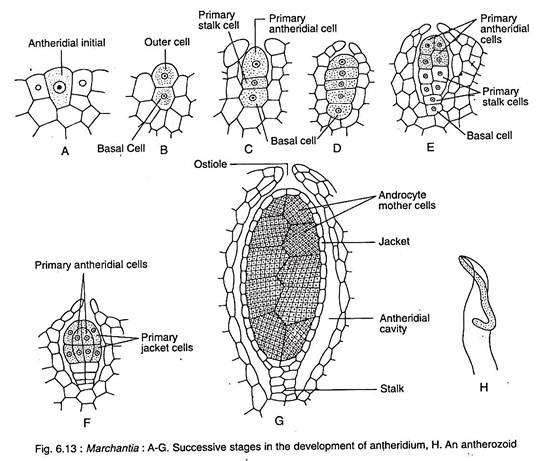
The development of antheridium in Marchantia is similar to that of Riccia. The antheridium develops from the antheridial initial, situated on the dorsal surface of the disc of the antheridiophore. The antheridial initial increases in size and a transverse division differentiates it to an outer cell and a basal cell (Fig. 6.1 3A, B).
The basal cell follows no further development. The outer cell later undergoes transverse division forming a basal primary stalk cell and a terminal primary antheridial cell (Fig. 6.13C). Eventually the antheridium is formed from the primary antheridial cell. The basal primary stalk cell however, forms the stalk of the antheridium (Fig. 6.13D-G).
A mature antheridium has a short stalk and a globular body encircled by a sterile jacket which encloses a number of sperm mother cells. A single sperm mother cell forms two triangular androcytes, each of which develops a biciliated sperm or antherozoid (Fig. 6.13H). The dehiscence of antheridium in Marchantia is similar to that of Riccia.
ADVERTISEMENTS:
Archegoniophore or Carpocephalum:
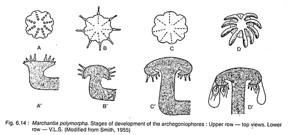
The archegoniophore or the carpocephalum is comprised of a stalk and a disc that bears 9 rays (Fig. 6.14D) instead of lobes. In the early stage, the archegonia develop on the upper (dorsal) side of the disc (Fig. 6.14A, A’). About 12-14 archegonia are arranged in a single row on each ray of the disc.
The stalk of the archegoniophore begins to elongate just after fertilisation and the central sterile part of the disc starts to enlarge enormously. As a result, the marginal apical region of the disc containing archegonia are pushed down to the lower-side of the disc (Fig. 6.14B, B’).
ADVERTISEMENTS:
The archegonia are hanging upside down — the youngest one being nearer the stalk while the older ones towards the periphery (Fig. 6.14C, C’). Subsequently, the tissue in between the rows of archegonia develops and hang down as rays. Initially the number of lobes is 8, later 9 rays are formed by the splitting of one.
Rays are long, stout and green finger-like projections that give the mature female receptacle an umbrella-like appearance (Fig. 6.14D, D’). The archegonia become inverted by the curvature of the disc. Single layered plate of tissue overlaps on either side of row of archegonia to form perichaetium or involucre. This plate of tissue encloses all the archegonia (about 12 to 14) arranged in a single row (Fig. 6.15A).
Archegonium:
Development and Structure of Archegonium:
Like Riccia, the archegonia of Marchantia also develop from a single superficial dorsal cell, called the archegonial initial (Fig. 6.16A). The conical shaped initial cell becomes conspicuous, increases in size, and projects above the surface.
ADVERTISEMENTS:
The initial cell divides by a transverse wall to form an upper (or outer) primary archegonial cell and a lower (or inner) primary stalk cell (Fig. 6.16B). The primary stalk cell by a few irregular divisions forms a short but distinct multicellular stalk of the archegonium. The primary archegonial initial undergoes several phases of regular divisions to form the archegonium (Figs. 6.16B-H).
A mature archegonium (Figs. 6.15B) is a pendulous flask-shaped structure with a short stalk, which attaches to the lower (ventral) surface of the archegonial disc. A single archegonium has a basal swollen venter containing an egg cell, a ventral-canal cell and a row of neck canal cells inside the elongated neck.
Fertilisation of Archegonium:
Like other bryophytes, the fertilisation of Marchantia is dependent upon the presence of water. Marchantia is dioecious or heterothallic, so the fertilisation is possible only when the male and the female plants grow together. The process of fertilisation takes place at a stage when the archegonia are in upright position on the upper surface of the disc of the very short archegoniophore.
The antherozoids of the antheridiophore discs are splashed by rain water on to the archegoniophores. The rest of the procedure of the fertilisation is the same as in Riccia. The archegoniophore is further developed in size after fertilisation which eventually leads to the inversion i.e., upside down position of the archegonia. In this phase the development of rays and perichaetium take place.
Sporophyte:
Development of the Sporophyte:
The fertilised egg enlarges in size and fills up the cavity of the venter. It invests itself immediately with a thin cellulose wall and becomes the oospore or zygote which is the mother cell of the sporophytic generation.
The zygote cell inside the venter divides first by a transverse wall forming an outer epibasal cell and an inner hypobasal cell (Fig. 6.17A). This is followed by a second division at right angle to the first division forming a quadrant of cells. The two upper epibasal cells of the quadrant divide and re-divide to form the capsule and the upper part of the seta.
The two hypobasal cells after several divisions form the lower part of the seta and the foot. The cells within the capsule are early differentiated into the peripheral single layered amphithecium and the central many- celled endothecium (Fig. 6.17C). A single- layered outer jacket is formed from the anticlinal divisions of amphithecial cells.
All the cells of endothecium on the other hand functions as archesporium and forms a massive sporogenous tissue. Subsequently, about half of the sporogenous tissues by repeated divisions, form a large number of spore mother cells. Each spore mother cell following a meiotic division gives rise to a spore tetrad.
The remaining cells of the sporogenous tissue function as elater mother cells and, subsequently, become elongated, endowed with two spiral thickenings and become the sterile elaters (Fig. 6.18A, D). Unlike spores, the elaters are diploid cells and are hygroscopic, help in spore dispersal.
The early development of zygote is associated with several significant changes in the gametophytic tissue surrounding the embryo. The stimulus of fertilisation leads to-the periclinal division of the venter wall which eventually gives rise to a 2-3 layered calyptra enveloping the growing sporangium. A ring of basal cells of archesporium forms a second one cell thick protective covering round the growing sporangium.
It is called the perigynium or the pseudoperianth. Therefore, the sporangium of Marchantia develops within three gametophytic coverings viz., calyptra, perigynium and perichaetium (Fig. 6.18A-C).
Structure of the Mature Sporophyte:
The mature sporophyte of Marchantia (Fig. 6.18A) is differentiated into three parts — foot, seta and capsule.
(a) Foot:
It is an expanded bulbous mass of cells at the base of the sporogonium, serves as an absorbing and anchoring organ. It absorbs nutrients from the gametophyte.
(b) Seta:
It is a slightly elongated stalk that connects the foot to the capsule. The cells are parenchymatous and elongated lengthwise. Thus, seta increases in length and pushes the mature capsule out of the calyptra.
(c) Capsule:
It is a yellow-coloured spherical structure, contains numerous spores and elaters. The capsule is protected by three coverings viz. calyptra perigynium and perichaetium.
Dehiscence of the Capsule:
The jacket of the hanging mature sporogonium (capsule) gets dried and splits longitudinally into a variable number of longitudinal lobes (Fig. 6.18B). These lobes extend from the apex to the middle of the capsule. The discharge and dispersal of sport to the air are assisted by the coiling and uncoiling of the elaters due to their hygroscopic nature.
The spores are almost rounded and are very minute in size (12 to 30 µm in diameter) with a thin inner intine and a slightly thicker outer exine (Fig. 6.18D). In some species an additional layer called perispore is also found outside the exine.
The new Gametophyte:
Spore is the first cell of the gametophyte which germinates to form a new gametophytic plant (Fig. 6.19A). Spores remain viable for about a year. Under favourable conditions they absorb moisture from the substratum and increase in size (Fig. 6.19B).
At this stage chloroplasts reappear. Subsequently, the spore divides transversely to form two unequal cells (Fig. 6.19C). The smaller cell forms the first rhizoid, while the larger cell undergoes irregular divisions to form a filamentous structure (Fig. 6.19D-E) which ultimately forms the normal Marchantia thallus.
Marchantia is a dioecious plant. Out of each spore tetrad, two spores grow into two male plants and the other two into two female plants. Fig. 6.20 represents the life cycle of Marchantia.


