ADVERTISEMENTS:
In this article we will discuss about:- 1. Habit and Habitat of Nephrolepis 2. External Features of Nephrolepis 3. Internal Structure 4. Reproduction.
Habit and Habitat of Nephrolepis:
Nephrolepis (Nephros, kidney; lepis, the indusium kidney-shaped and scale like) is represented by about 30 species. These species are distributed in the tropics of the entire world.
About five species [N.cordifolia (Fish bornfern, Narrow sword fern), N. exaltata (sword fern), N. volubilis, N. acuta and N. ramosa] are found in India. Majority of the species are terrestrial but a few species e.g., N. ramosa are found climbing on the trees. N. cordifolia is found throughout the Indian region upto 5000 feet elevation.
External Features of Nephrolepis:
ADVERTISEMENTS:
The mature plant body is sporophytic and can be differentiated into rhizome, roots and leaves.
1. Rhizome:
It is short, slender, suberect (TV. cordifolia), erect (N. exaltata), or wide creeping (N. volubilis, goes upto 50 feet over trees). It bears a close tuft of leaves and long, slender lateral branches called runners. The runners spread for a considerable distance and bear roots (Fig. 1).
2. Roots:
ADVERTISEMENTS:
Branched adventitious roots arise from the rhizome and runners in aeropetal succession.
3. Leaves:
Leaves are tufted, long, narrow and simply pinnate. Steple (a leaf stalk) is 2.5 to 10 cm long in N. cordifolia and upto 20 cm long in N. acuta. Fronds (leaves Fig. 2 A, B) are 30 cm (N. ramosa) to 240 cm long (N. acuta), cm long (N. acuta) Pinnae are numerous, crowded, often imbricated, 3 cm long (N. cordifolia) to 20 cm long (N. acuta), slightly falcate and articulated at the base.
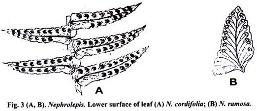
Margins are entire or slightly crenate. The veins are free and there are present white line dots over the vein tips. Sori are present on the lower surface. Sori are half the way between the midrib and margin in a single row [(N. cordifolia), (Fig. 3 A, B)] or submarginal (N. exaltata) or near the margin (N. acuta).
The indusia are usually round-reniform with a narrow sinus. Sometimes the sinus widens to a broad curved base. Young leaves are covered with multicellular hairs and scales and show circinate vernation.
Internal Structure of Nephrolepis:
1. Transverse Section (T.S.) of Rhozome:
Internal structure of rhizome can be differentiated into three parts: epidermis, cortex and stele. Epidermis is outermost layer. It is made up single layer of narrower cells. Epidermis is followed by cortex. Stele is primitive type of dictyostele. Two curved strands facing each other are present in the centre. Both strands are separated from each other and are surrounded by sclerenchyma. Each strand has its own pericycle and endodermis (Fig. 4).
2. Transverse Section of Runner:
Internally it is also differentiated into epidermis, cortex and stele (Fig. 5). The outermost layer is epidermis. It has stomata at young stages.
Epidermis is followed by a few layers of closely packed cells (intercellular spaces are absent). The remaining layers are made up of large, round or oval cells with abundant intercellular spaces. Cortex is followed by endodermis. Just inside the endodermis lies the pericycle.
ADVERTISEMENTS:
Stele consists Xylem cylinder in the form of fluted column, composed of tracheids and parenchyma. The protoxylem consists of 7-9 distinct exarch strands forming the ridges of the xylem cylinder. Phloem goes round the xylem. It is many cells thick in the bays between protoxylem ridges and also forms a wavy mantle over the protoxylem ridges.
Anatomy of the Leaf:
3. Transverse Section of Petiole:
The petiole has an adaxial groove. The internal structure of petiole can be differentiated into three parts: epidermis, cortex and stele (Fig. 6). Epidermis is outermost protective layer. It is composed of small thick walled cells.
ADVERTISEMENTS:
Epidermis is followed by 3-4 layers of sclerenchymatous cortex and compact parenchyma. Usually 3-6 conducting strands are embedded in the parenchymatous cortex and are arranged in horse-shoe like shape.
The xylem portion of each strand is crecentric. Protoxylem lies towards outside. Phloem completely surrounds xylem which in turn is surrounded by a layer of pericycle and endodermis.
4. Transverse Section of Pinnule or Lamina:
ADVERTISEMENTS:
It is similar to Pteridium. The transverse section of the sporophyll passing through sori reveals that shield shaped indusium is attached to the receptacle. Club shaped stalked sporangia are attached, to the receptacleand are protected by indusium.
5. Transverse Section of Root:
It is circular is outline and can be differentiated into epidermis, cortex and stele. Epidermis is single layered. Cortex is wide, many layered and parenchymatous. The stele is usually diarch and exarch. Xylem is surrounded by pericycle and endodermis each.
Reproduction in Nephrolepis:
Nephrolepis reproduces by two methods:
(i) Vegetative reproduction.
(ii) Sexual reproduction.
ADVERTISEMENTS:
(i) Vegetative reproduction:
It takes place by the formation of buds. Runner produces buds at some distance from parent rhizome. These buds give rise to fronds and thus help in vegetative reproduction of the plants.
ADVERTISEMENTS:
(ii) Sexual Reproduction:
Structure and Development of Sporangium:
Structure and development of sporangium is similar to Pteridium. Mature spores are brown in colour, with thin exosporium, thick endosporium with small warts.
Structure and Development of Prothallus:
Development of prothallus is similar to Pteridium. A mature prothallus is formed in about fifty days. It is large, green and heart shaped (Fig. 7) and 0.3 cm x 0.5 cm in diameter. It has a large cushion on the ventral side. It is 4 – 6 cells thick in the region of cushion and one celled elsewhere.
It has a deep notch and 4-7 meristematic cells lie in it. Prothallus is monoecious. At maturity antheridia are first to appear anteroposteriorly on the posterior side of the cushion. Development and structure of antheridium is similar to Pteridium.
A mature antheridium is large and spherical. At maturity it consists of 32 spermatocytes. Structure and development of the archegonium is similar to Pteridium. Many archegonia are formed and lie in the anterior part of the cushion at maturity. Fertilization and development of sporophyte is also similar to Pteridium.


