ADVERTISEMENTS:
In this article we will discuss about Fasciola Hepatica:- 1. Habit and Habitat of Fasciola Hepatica 2. Structure of Fasciola Hepatica 3. Body Wall 4. Digestive System 5. Respiration 6. Excretion 7. Nervous System 8. Reproductive System 9. Life History 10. Parasitic Adaptations.
Contents:
- Habit and Habitat of Fasciola Hepatica
- Structure of Fasciola Hepatica
- Body Wall of Fasciola Hepatica
- Digestive System of Fasciola Hepatica
- Respiration in Fasciola Hepatica
- Excretion in Fasciola Hepatica
- Nervous System of Fasciola Hepatica
- Reproductive System of Fasciola Hepatica
- Life History of Fasciola Hepatica
- Parasitic Adaptations of Fasciola Hepatica
1. Habit and Habitat of Fasciola Hepatica:
Fasciola hepatica (L., fasciola = small bandage; Gr., hepar = liver), the sheep liver fluke, lives as an endoparasite in the bile passages of sheep.
ADVERTISEMENTS:
Its life cycle is digenetic, i.e., completed in two hosts (a primary vertebrate host, the sheep and a secondary or intermediate invertebrate host, the gastropod mollusc). The adult parasite is found in the primary host, while a part of its life cycle as larval stages are found in the invertebrate host.
Fasciola hepatica, in addition to sheep, also infects other vertebrates like goat, deer, horse, dog, ass, ox and occasionally man. Its secondary hosts are either Planorbis sps, Bulinus sps., or Limnaea truncatula, all being freshwater gastropod molluscs. Fasciola hepatica is worldwide in distribution, particularly sheep and cattle raising areas are the primary zones where human beings are also infected.
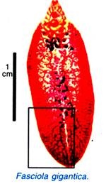
Its other Indian species, F. gigantica (= indica) is found in the bile passages of buffaloes, cow, goats and pigs.
2. Structure of Fasciola Hepatica:
(i) Shape, Size and Colour:
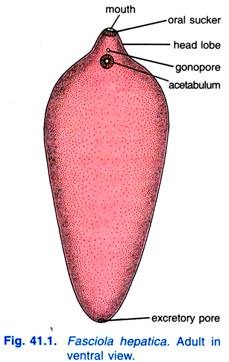
F. hepatica has a thin, dorsoventrally flattened, leaf-shaped, elongated and oval body. It measures about 25 to 30 mm in length and 4 to 12 mm in breadth.
The maximum width is at about anterior third of the body from where the body tapers anteriorly as well as posteriorly, however, the anterior end is somewhat rounded, while it is bluntly pointed posteriorly. F. indica has its greatest width at about the middle of the body, and the posterior end is rounded. It is usually pinkish in colour but it appears brownish due to ingested bile of the host.
(ii) External Morphology:
The anterior end of the body is distinguished into a triangular oral cone or head lobe giving it a shouldered appearance. The head lobe, at its tip, bears a somewhat triangular aperture called mouth. There are two muscular suckers an oral sucker at the anterior end encircling the mouth, and a large ventral sucker or acetabulum situated mid-ventrally about 3 to 4 mm behind the oral sucker.
The suckers are cup-like muscular organs meant for attachment to the host by vacuum. In addition to mouth aperture, there are two permanent apertures on the body; one situated mid-ventrally in front of the ventral sucker is the common genital aperture or gonopore, and the other is situated at the posterior end of the body called the excretory pore.
In addition to these apertures, a temporary opening of Laurer’s canal appears during the breeding season on the dorsal surface just anterior to the middle of the body. Anus is wanting because alimentary canal is incomplete.
3. Body Wall of Fasciola Hepatica:
The body wall of F. hepatica lacks a cellular layer of epidermis, unlike those of the turbellarians. However, it consists of a thick layer of cuticle followed by a thin basement membrane and underlying muscle layers surrounding the mesenchyma.
(i) Cuticle:
A tough resistant cuticle, made of a homogeneous layer of scleroprotein, covers the fluke and protects it from the juices of the host. It bears small spines, spinules or scales. The spinules anchor the fluke to the bile duct of the host, provide protection and facilitate locomotion.
ADVERTISEMENTS:
The cuticle of F. indica has broad, stout, and blunt scales. The epidermis has been lost during development of the cercaria stage. However, the cuticle is secreted by special mesenchymal cells situated below muscle layers. These cuticle secreting cells were believed to be sunken epidermal cells (Hein, 1904 and Roewer, 1906).
(ii) Basement Membrane:
The lowest layer of the cuticle is a thin, delicate basement membrane. It demarcates the boundary between cuticle and muscle layers.
(iii) Muscle Layer:
ADVERTISEMENTS:
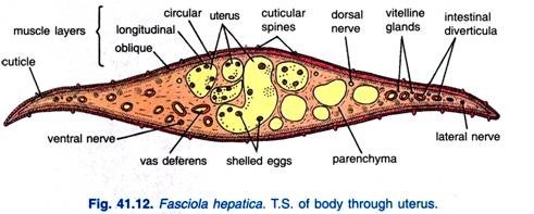
The basement membrane is followed by a sub-cuticular musculature. It consists of an outer layer of circular muscle fibres, middle layer of longitudinal muscle fibres and an inner layer of diagonal muscle fibres which are more developed in the anterior half of the body. All muscles are smooth. The muscles form stout bundles of radial fibres in the suckers.
(iv) Mesenchyme:
Below the muscles is parenchyma (mesenchyme) having numerous loosely arranged uninucleate and bi-nucleate cells with syncytial network of fibres having fluid-filled spaces.
Some of these cells are large and provided with large processes extending up to the base of the cuticle to which they are said to secrete. In fact, the mesenchyme forms a packing material between the muscle layer and internal organs. It helps in the transport of nutrients and waste substances.
The body wall plays a significant role in the physiology of fluke. It provides protection, it is the site of gaseous exchange, various nitrogenous wastes are diffused out through it and it also helps in the absorption of amino acids to some extent.
(v) Structure of Body Wall Under Electron Microscope:
ADVERTISEMENTS:
The electron microscopic studies of the body wall of F. hepatica by Threadgold (1963), Bils and Martin (1966) have clearly revealed that cuticle is a syncytial layer of protoplasm having mitochondria, endoplasmic canals, vacuoles and pinocytic vesicles. Hence, cuticle is now referred to as the integument because it is metabolically active. The tegument is continuous with tegument secreting cells lying in the mesenchyme.
The outer integumental surface is thrown out into many fine projections which increase its area to facilitate the absorption of host’s fluid. The tegument is also provided with many fine pore canals through which dissolved substances in the form of solution are absorbed into the mesenchyme.
4. Digestive System of Fasciola Hepatica:
(i) Alimentary Canal:
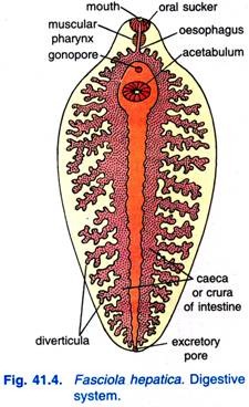
The oral sucker encloses a ventral mouth which leads into a funnel- shaped mouth cavity, followed by a round muscular pharynx with thick walls, and a small lumen. The pharynx has pharyngeal glands. F. indica has a short muscular pharynx from which arises an oral pouch which is about half the size of the pharynx.
ADVERTISEMENTS:
There is a short narrow oesophagus leading into an intestine which divides into two branches or intestinal caeca or crura each running on one side to the posterior end, and ending blindly. The intestinal caeca give out a number of branching diverticula in order to carry food to all parts of the body since there is no circulatory system. The median diverticula are short and lateral ones are long and branching. There is no anus.
The interior part of the alimentary canal up to the oesophagus is lined with cuticle and serves as a suctorial fore gut; the intestine is lined with endodermal columnar epithelial cells. The caceal epithelium has secretory gland cells.
(ii) Food, Feeding and Digestion:
It feeds on bile, blood, lymph and cell debris. The oral sucker and pharynx together constitute an effective suctorial apparatus. Digestion is extracellular, occurs in intestine. The digested food material is distributed by branching diverticula of intestine to all parts of the body as the circulatory system is not found in this animal. Thus, the digestive system functions as a gastro vascular system.
In fact, the digested nutrients are passed into the parenchyma through intestinal diverticula; from parenchyma they are diffused into the various organs of the body.
Reserve food, mostly in the form of glycogen and fats is stored in the parenchyma. However, monosaccharide sugars like glucose, fructose, etc., are directly diffused into the body of the fluke through general body surface from the surrounding fluid of the host. The indigestible remains of the food, if any, are probably said to be ejected through the mouth.
5. Respiration of Fasciola Hepatica:
Mode of respiration is anaerobic or anoxybiotic. In fact, glycogen is metabolised to carbon dioxide and fatty acids releasing energy in the form of heat.
ADVERTISEMENTS:
The process is completed in following steps:
(i) The glycogen undergoes anaerobic glycolysis to form pyruvic acid,
(ii) The pyruvic acid is decarboxylated to form carbon dioxide and an acetyl group,
(iii) The acetyl group then combines with coenzyme A to form acetyl coenzyme A, and
(iv) The acetyl coenzyme A is then finally condensed and reduces to form fatty acids.
ADVERTISEMENTS:
The carbon dioxide, thus, produced is diffused out through general body surface and the fatty acids are excreted through the excretory system.
6. Excretory System of Fasciola Hepatica:
The excretory system of Fasciola hepatica is concerned with excretion as well as osmoregulation. It consists of a large number of flame cells or flame bulbs or protonephridia connected with a system of excretory ducts.
(i) Flame Cells:
The flame cells, supposed to be modified mesenchymal cells, are numerous, irregular in shape bulb-like bodies found distributed in the mesenchyma throughout the body of Fasciola. The distribution pattern of flame cells follows a specific pattern referred to as ‘the flame cell pattern’ (Faust, 1919).
The flame cells are characteristic, each has a thin elastic wall with pseudopodia-like processes, a nucleus and an intracellular cavity having many long cilia arising from basal granules. In living condition, the cilia vibrate like a flickering flame, hence, the name flame cell.
(ii) Excretory Ducts:
There is an excretory pore at the posterior end from which arises a longitudinal excretory canal, from this arise four main branches, two dorsal and two ventral, which subdivide into numerous small capillaries which anastomose; the capillaries are continued into the intracellular cavity of flame cells. The longitudinal excretory canal is non-ciliated but the capillaries are lined with cilia.
(iii) Process of Excretion:
The excretory wastes, generally fatty acids and ammonia, are diffused from surrounding mesenchyma into the flame cells and finally collected into their intracellular cavities. The vibrating movement of the cilia causes the flow of wastes from the intracellular cavities of flame cells into the excretory ducts and then into the main excretory canal and finally to the exterior through excretory pore by hydrostatic pressure.
Such an excretory system of flame cells and canals or ducts of various orders with no internal opening and leading to an excretory pore which opens to the exterior is spoken of as a protonephridial system which is excretory but its main function is to regulate the amount of fluid in the animal’s body.
7. Nervous System of Fasciola Hepatica:
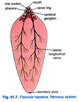
A nerve ring surrounds the oesophagus, it has a pair of cerebral ganglia dorsolaterally, and a ventral ganglion below the oesophagus. Small nerves are given out anteriorly from the ganglia. Posteriorly three pairs of longitudinal nerve cords arise from the ganglia, a dorsal, a lateral, and a ventral pair of nerve cords.
The lateral nerve cords are best developed and they run to the posterior end. Nerve cords are connected by transverse commissures and they give out many small branches, some of which form plexuses. The nerve cells are mostly bipolar. Due to parasitic life, sense organs are lost in adult Fasciola.
8. Reproductive System of Fasciola Hepatica:
Fasciola hepatica is hermaphrodite but usually cross fertilisation takes place. The reproductive organs are well developed and complex.
(i) Male Reproductive System of Fasciola Hepatica:
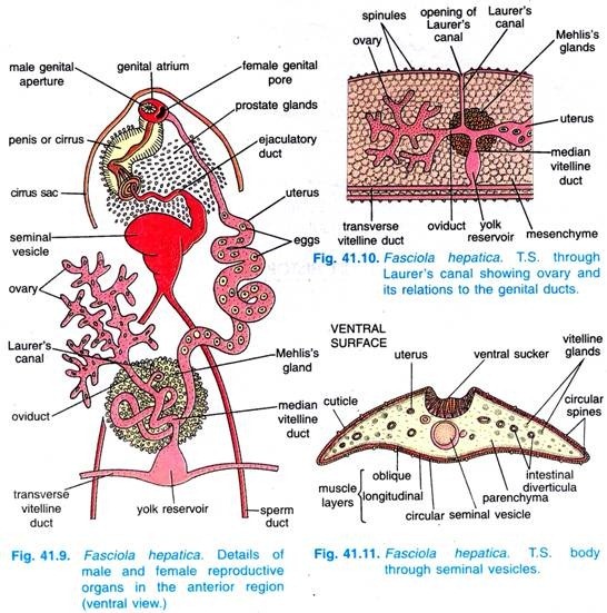
The male reproductive system consists of testes, vasa deferentia, seminal vesicle, ejaculatory duct, cirrus or penis, prostate glands and genital atrium.
(a) Testes:
These are two in number, much ramified tubular and placed one behind the other (i.e., with tandem arrangement) in the posterior middle part of the body. In fact, they occupy major space from behind the middle part of the body of Fasciola. The cells lining the wall of testes give rise to spermatozoa.
(b) Vasa Deferentia:
A narrow and slender vas deferens or sperm duct arises from each testis and runs forwards.
(c) Seminal Vesicle:
The two vasa deferentia unite together near the acetabulum (ventral sucker) and become dilated to form a muscular, elongated, broad, bag-like seminal vesicle or vesicula seminalis. It serves the purpose of storing sperms.
(d) Ejaculatory Duct:
The seminal vesicle continues anteriorly into a very narrow and coiled duct called ejaculatory duct.
(e) Cirrus:
The cirrus (penis) is a muscular and elongated structure into which ejaculatory duct opens. The cirrus opens by male genital aperture in a common genital atrium. The cirrus of F. indica is covered with small spines.
(f) Prostate Glands:
The ejaculatory duct is surrounded by numerous unicellular prostate glands. These glands open into the ejaculatory duct and their secretion (alkaline) helps in free movement of sperms during copulation.
(g) Genital Atrium:
The genital atrium is a common chamber for male and female genital apertures, it opens externally by a gonopore lying ventrally in front of the acetabulum. The cirrus can be everted through the gonopore during copulation. The cirrus or penis, seminal vesicle and prostatic glands are surrounded in a common cirrus sheath or cirrus sac.
(ii) Female Reproductive System of Fasciola Hepatica:
The female reproductive system consists of ovary, oviduct, uterus, vitelline glands, Mehlis’s glands and Laurer’s canal.
(a) Ovary:
The ovary is single, tubular, highly branched and situated to the anterior of testes at the right side in anterior one-third of the body.
(b) Oviduct:
All the branches of ovary open into a short and narrow tube called oviduct. The oviduct travels down obliquely and opens into the median vitelline duct.
(c) Uterus:
From the junction of oviduct and median vitelline duct arises a wide convoluted uterus having fertilised shelled eggs or capsules. The uterus opens by female genital aperture into the common genital atrium on the left side of male genital aperture. The uterus is comparatively small and it lies in front of the gonads.
The terminal part of uterus has muscular walls, referred to as metraterm which ejects the eggs and also sometimes receives the cirrus during copulation.
(d) Vitelline Glands:
On both lateral sides and also behind the testes are numerous follicles constituting the vilellaria, yolk glands or vitelline glands which produce albuminous yolk and shell material for the eggs. The vitelline glands open by means of minute ducts into a longitudinal vitelline duct on each side.
The two longitudinal ducts are connected together by a transverse vitelline duct placed above the middle of the body. The transverse vitelline duct is swollen in the centre to form the yolk reservoir or vitelline reservoir. From the yolk reservoir a median vitelline duct starts and runs forward to join the oviduct.
(e) Mehlis’s Glands:
A mass of numerous unicellular Mehlis’s glands is found situated around the junction of median vitelline duct, oviduct and uterus. The secretion of Mehlis’s glands lubricates the passage of eggs in the uterus and probably hardens the egg shells, it probably also activates spermatozoa.
The junction of oviduct and median vitelline duct is swollen to form ootype in certain flukes like F. indica, in which the parts of an egg are assembled and the eggs are shaped, but an ootype is lacking in F. hepatica (according to some authorities).
(f) Laurer’s Canal:
From the oviduct arises a narrow Laurer’s canal, it runs vertically upwards. This canal opens on the dorsal side temporarily during breeding season and acts as vestigial vagina to serve as copulation canal.
9. Life History of Fasciola Hepatica:
(i) Copulation and Fertilization of Fasciola Hepatica:
Though F. hepatica is hermaphrodite even then cross- fertilisation is of common occurrence. Hence, before fertilisation copulation occurs; during copulation, which occurs in the bile duct of the sheep, the Cirrus of one Fasciola is inserted into the Laurer’s canal of other Fasciola and the sperms are deposited into the oviduct, so that cross-fertilisation takes place.
During self- fertilisation, which occurs only when cross-fertilisation does not take place, the sperms from the same Fasciola enter the female genital aperture and pass down the uterus to fertilize the eggs in the oviduct.
(ii) Formation of Egg Capsules in Fasciola Hepatica:
The eggs are brownish in colour, oval in shape and measure about 130 to 150 µ in length and 63 to 90 µ in width.
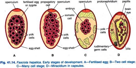
As referred to, the eggs are fertilised in the oviduct, the fertilised eggs receive yolk cells from vitelline glands and they get enclosed in a chitinous shell formed by granules in the yolk cells giving out droplets, the shell hardens and becomes brownish yellow; the shell has an operculum or lid. Mehlis’s glands play no role in the formation of the shell.
The completed ‘eggs’ are called capsules which are large in size and they pass into the uterus where development starts. Capsules come out of the gonopore into the bile duct of the sheep, they reach the intestine and are passed out with the faeces. The capsules which fall in water or damp places will develop at about 75°F. Capsules are produced throughout the year, and one fluke may produce 500,000 capsules.
(iii) Development of Fasciola Hepatica:
Development starts in the uterus and is continued on the ground. The fertilised egg divides into a small propagatory cell and a larger somatic cell. The somatic cell divides and forms the ectoderm of the larva. Later the propagatory cell divides into two cells, one of which forms the endoderm and mesoderm of the larva, and the other forms a mass of germ cells at the posterior end of the larva.
This method of development takes place in the formation of all larval stages during the life history. In two weeks time, a small ciliated miracidium larva is formed and it comes out of the shell by forcing the operculum. The miracidium produces a proteolytic enzyme which erodes the lower surface of the operculum.
(iv) Miracidium Larva:
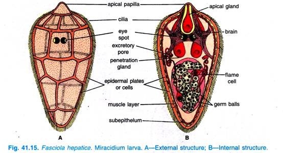
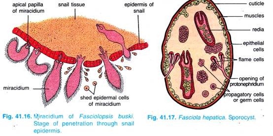
Miracidium larva is a minute, oval and elongated, free-swimming stage, it is covered with 18 to 21 flat ciliated epidermal cells lying in five rings.
The first ring is made of six plates (two dorsal, two lateral and two ventral), second ring has again six plates (three dorsal and three ventral), third ring has three plates (one dorsal and two ventrolateral), fourth ring has four plates (two right and two left) and fifth ring has two plates (one left and one right).
A sub-epidermal musculature, consisting of outer circular and inner longitudinal fibres, is situated beneath the epidermal cells. The sub-epidermal musculature is followed by a layer of cells constituting the sub-epithelium. All these, i.e., epidermal cells, sub-musculature and sub- epithelium, together form the body wall of miracidium.
Anteriorly it has a conical apical papilla, and attached to it is a glandular sac with an opening called apical gland.
On each side of the apical gland is a bag-like penetration gland. There are two pigmented X-shaped eye spots and a nervous system. There is a pair of protonephridia, each with two flame cells. The flame cells open to the exterior by two separate excretory pores or nephridiopores situated laterally in the posterior half of the body.
Towards the posterior side are some propagatory cells (germ cells), some of which may have divided to form germ balls which are developing embryos. The miracidium does not feed, it swims about in water or moisture film, but it dies in eight hours unless it can reach a suitable intermediate host, which is some species of amphibious snail of genus Limnaea or even Bulinus or Planorbis.
After getting a suitable host the miracidium adheres to it by its apical papilla and enters the pulmonary sac of the snail, from where it penetrates into the body tissues with the aid of penetration glands and finally reaches to snail’s digestive gland. In the tissues the miracidium casts off its ciliated epidermis, loses its sense organs and it swells up and changes in shape to form a sporocyst.
(v) Sporocyst:
The sporocyst is an elongated germinal sac about 0.7 mm long and covered with a thin cuticle, below which are mesenchyme cells and some muscles.
The glands, nerve tissue, apical papilla and eye spots of miracidium disappear. The hollow interior of sporocyst has a pair of protonephridia each with two flame cells it has germ cells and germ balls. The germ cells have descended in a direct line from the original ovum from the miracidium developed.
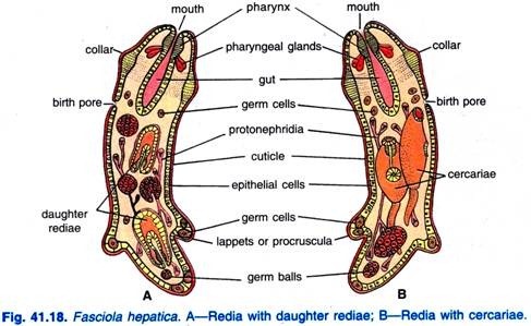
The sporocyst moves about in the host tissues and its germ cells develop into a third type of larva called redia larva. A sporocyst forms 5 to 8 rediae. The rediae larvae pass out of the sporocyst by rupture of its body wall into the snail tissues with the aid of the muscular collar and ventral processes, then the rediae migrate to the liver of the snail.
(vi) Redia:
The redia is elongated about 1.3 mm to 1.6 mm in length with two ventral processes called lappets or procruscula near the posterior end and a birth pore near the anterior end.
Body wall has cuticle, mesenchyme and muscles, and near the anterior end, just in front of the birth pore, the muscles form a circular ridge, the collar used for locomotion. Redia has an anterior mouth, pharynx in which numerous pharyngeal glands open, sac-like intestine and there is a pair of protonephridia with two pairs of flame cells. Its cavity has germ cells and germ balls.
The germ cells of redia give rise during summer months to a second generation of daughter rediae, but in winter they produce the fourth larval stage, the cercaria larva. Thus, either the primary redia or daughter redia produce cercaria larvae which escape from the birth pore of the redia into the snail tissues. Each redia forms about 14 to 20 cercariae.
(vii) Cercaria:
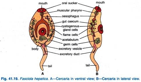
The cercaria (Fig. 41.19) has an oval body about 0.25 mm to 0.35 mm long and a simple long tail. Its epidermis is soon shed and replaced by cuticle; below the cuticle are muscles and cystogenous glands. It has rudiments of organs of an adult; there are two suckers (oral sucker and ventral sucker) and an alimentary canal consisting of mouth, buccal cavity, pharynx, oesophagus and a bifurcated intestine.
There is an excretory bladder with a pair of protonephridial canals (excretory tubules) with a number of flame cells. An excretory duct originates from the bladder, travels through the tail and bifurcates to open out through a pair of nephridiopores.
There are two large penetration glands, but they are non-functional in the cercaria of Fasciola.
It also has the rudiments of reproductive organs formed from germ cells. The cercariae escape from the birth pore of the redia, then migrate from the digestive gland of the snail into the pulmonary sac from where they pass out into surrounding water. The time taken in snail from the entry of miracidia to the exit of cercariae is five to six weeks.
(viii) Metacercaria:
The cercariae swim about in water for 2 to 3 days; they then lose their tails and get enclosed in a cyst secreted by cystogenous glands.
The encysted cercaria is called a metacercaria (Fig. 41.20) which is about 0.2 mm in diameter and it is in fact a juvenile fluke. If the metacercariae are formed in water they can live for a year, but if they are formed on grass or vegetation then they survive only for a few weeks, they can withstand short periods of drying.
The various larval stages (the miracidium, sporocyst, redia, and cercaria) are all formed in the same way from germ cells which are set aside at the first division. There is, thus, a distinction between germ cells and somatic cells, and germ cells alone form the various larval stages.
Infection of the primary host (Sheep):
Further development of the metacercaria takes place only if it is swallowed by the final host, the sheep.
Metacercariae can also infect man if they are swallowed by eating water cress on which cercariae encyst, but such cases are rare. But the metacercarie are not infective until 12 hours after encystment. In the alimentary canal of a sheep, the cyst wall is digested and a young fluke emerges and bores through the wall of the intestine to enter the body of the host.
After about two to six days they enter the liver and their movements in the liver may cause serious injuries.
The young flukes stay in the liver for seven or eight weeks feeding mainly on blood and then they enter the bile duct and bile passages. The young flukes have been growing in the liver and after several weeks in the bile duct they become sexually mature adults. The period of incubation in the sheep takes 3 to 4 months.
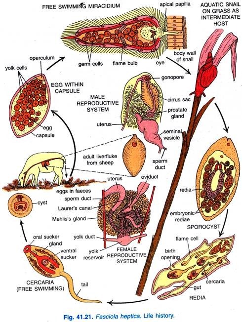
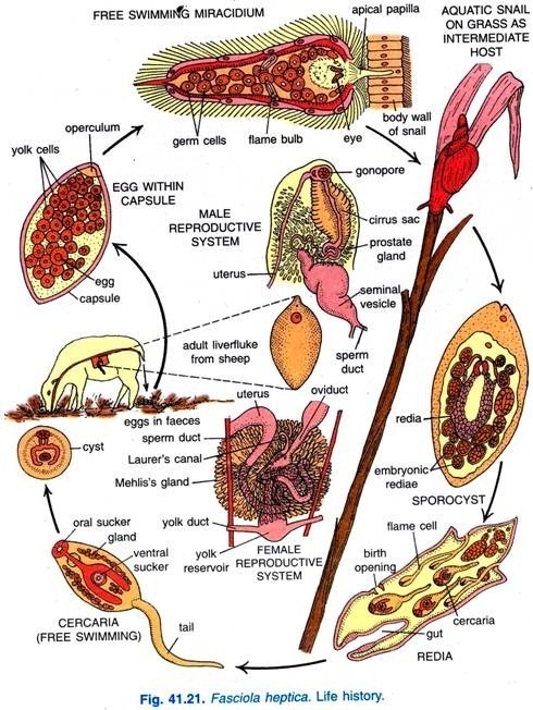
However, the life history of Fasciola hepatica (Fig. 41.21) can be summarised as under:
Adult flukes in liver → copulation and fertilisation → laying of capsules in the bile ducts → capsules in the intestine (stages in sheep’s body) → capsules out in faeces → miracidia escape from capsules (stages in open) → miracidia → sporocysts → rediae → cercariae → (stages in snail’s body) → cercariae → metacercariae (stages in open) → metacercariae young flukes → adult flukes (stages in a fresh sheep’s body).
Characteristic Features of Life History of Fasciola Hepatica:
Life history of Fasciola Hepatica is complicated because of parasitism. A sheep harbours about 200 flukes which will produce about 100 million eggs. The miracidium larva is free living and is structurally adapted to seek out an intermediate host, a snail Limnaea, which is found conveniently in water and damp places in grass in wide areas where sheep graze.
The sporocyst forms 5 to 8 rediae, each of which produces 8 to 12 daughter rediae, each daughter redia forms 14 to 20 cercariae; so that about a thousand cercaria larvae are produced from each egg. From this large number some cercariae are bound to infect a new sheep, thus, ensuring a continuance of the race.
Life history of Fasciola Hepatica affords an example of alternation of generations. The fluke is the sexual generation and it alternates, not with an asexual generation, but with parthenogenetic generations of sporocysts and rediae. Such an alternation of a sexual generation with a series of parthenogenetic generations is called heterogamy.
This theory of parthenogenetic development in the various larval stages is now discounted, and formation of various larvae from germ cells is regarded as simple mitotic asexual multiplication; this asexual multiplication of various larvae is called polyembryony.
Thus, there is a period of asexual multiplication during larval stages, followed by sexual reproduction in the adult fluke. This may be regarded as an alternation of generations, but more probably it is continuous life history in which asexual multiplication occurs in the larval stages due to parasitism.
The free swimming larval stages, miracidia and cercariae of F. hepatica, are morphologically more advanced than the adult fluke because they bear organs of locomotion, sense organs, cellular epidermis and a well developed body cavity.
10. Parasitic Adaptations of Fasciola Hepatica:
As adult Fasciola hepatica lives in the liver and bile ducts of sheep as an endoparasite, it is very well adapted for its parasitic mode of life. In fact, on one hand adult fluke exhibits certain adaptive features and on the other a number of adaptive features may also be accounted from the various stages of its life history.
So, the adaptive features of Fasciola hepatica can be discussed in the following two headings:
A. Adaptations of the Adult Fluke:
These can be accounted as under:
1. Its body is dorsoventrally flattened, leaf-shaped which increases surface area of the body for increased diffusion of substances through fluid of the body.
2. Its body is covered with a thick cuticle which protects it from host’s antitoxins.
3. Cilia are absent in adult flukes.
4. Adhesive organs like suckers (anterior sucker and ventral sucker) well developed which provide it firm attachment with the host tissue. Many cuticular spines over its body erode the host tissue forming its food and also serve in saving the fluke from being pushed away in the ducts with bile.
5. Its mouth is situated anteriorly and the muscular pharynx serves for sucking the nutrients from the host body. Since it feeds on pre-digested and digested substances of the host body, hence, its alimentary canal is not well developed and digestive glands are not found.
6. Since process of digestion does not occur, anus is absent and, hence, circulatory system is wanting because the various organs of alimentary canal (intestine and its various branches) distribute the already digested food substances to the different parts of its body.
7. Since it lives in an environment which is devoid of oxygen, hence, anaerobic mode of respiration occurs; respiratory organs are completely wanting.
8. Its nervous system is very simple and the sense organs are completely wanting, as the flukes are endoparasites.
9. Locomotory organs are not found as the flukes lead a well protected life.
10. The excretory system consists of a complicated arrangement of branched tubules so as to facilitate the collection of various metabolic excretory wastes of the body.
11. The reproductive system is well developed and best suited for its parasitic mode of life.
12. Since adult fluke lives in the body of the sheep, hence, it may die with the death of the sheep. Therefore, there is a need of secondary host for the transference of the parasite from one host to the other, so that its race may be continued. Hence, snail is the secondary host.
B. Adaptations in Life History:
The various parasitic adaptive features in the life history of Fasciola hepatica can be accounted as under:
1. Production of enormous number of eggs to overcome their wastage during transference.
2. The eggs are to pass down the bile duct into the intestine of sheep and then to the outside with its faeces, hence, the fertilised eggs are enclosed in a chitinous covering, the shell, which protects the zygote from the enzymes of the host. The shelled eggs are called capsules.
3. Miracidia are the first larvae to come out of the capsules; miracidia are well adapted to lead free swimming life (it has ciliated body to help in swimming, eye spots are developed) and also for entering into the body of the secondary host, Limnaea, Planorbis, etc. (penetration glands help them to enter into snail’s body).
4. The sporocyst leads parasitic life; its body is covered in a cyst-like structure to protect it from digestive enzymes of the snail. The germ balls in sporocyst give rise to rediae which may further produce either large number of daughter rediae or cercariae.
5. The locomotory organs of rediae (lappets or procruscula) and cercariae (tail) enable them to move and find their way into the fresh tissues of the snail.
6. Cercariae find their way out of the body of snail and lead a very short free life and then get enclosed in a cyst secreted by them itself on vegetations.
7. The cysted cercariae called metacercariae on vegetation make sure of their entry into the sheep’s body due to herbivorous habit of the sheep.
8. Metacercariae can live for a longer period waiting for entry into sheep’s body as they are well protected in cyst to overcome climatic hazards.
9. Mode of parthenogenetic reproduction of larvae further ensures the continuity of their race.
However, the high rate of reproduction, adaptations of the larvae, sexual and asexual mode of reproduction and adaptations in the morphology and physiology of the fluke are to ensure the survival and continuity of the race.
